Home Radiology and Imaging This service gives you the opportunity to perform radiology and medical imaging examinations in your home. It is provided to patients who have difficulty going to hospitals and clinics, and it includes various types of radiology.
-
Quick View
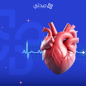
ECG imaging
₺4,500What is an electrocardiogram EKGEKG?
An electrocardiogram is also known as an electrocardiogram or EKG. It is a simple and quick test that records the heart’s electrical activity. It is used to assess the health of the heart. as well as to detect any other potential issues.
When is an EKG performed? EKG EKG?
- If you have symptoms that may indicate a heart condition. Like this: shortness of breath. chest pain. Irregular beats.
- If you have a medical condition that increases your risk of heart disease. Like this: diabetes. High blood pressure.
- When undergoing treatment for heart disease.
- During a routine physical exam.
What are the benefits of an EKG? EKG benefits?
- Diagnosis of heart disease. Like this: Irregular heartbeat heart attack. mansions.
- A general assessment of heart health.
- Monitor the effect of heart medications.
- Detecting future heart disease risk.
What to expect on an EKG EKG?
- Remove clothing and any jewelry that may interfere with the electrodes.
- Lie down at a comfortable table.
- The technician connects the electrodes to the body.
- An electrocardiogram is recorded for a few minutes.
- There is no pain or discomfort during the test.
-
Quick View
Echo imaging
₺9,000What is an echocardiogram?
An echocardiogram is a non-invasive ultrasound test that creates moving images of the heart and its valves. These images provide accurate and valuable information about the function and structure of the heart, helping to diagnose and evaluate many heart diseases.
When is an echocardiogram performed?
- When experiencing chest pain.
- When experiencing shortness of breath.
- When experiencing fatigue.
- When experiencing leg swelling.
- When experiencing difficulty exercising.
- When experiencing a chronic cough.
- When experiencing fainting.
- When experiencing an abnormal heart sound.
What are the benefits of an echocardiogram?
- Diagnosing heart diseases: An echocardiogram detects heart diseases including: valve diseases, pericardial diseases, cardiomyopathy, congenital defects, and blood clots.
- Evaluating heart function: An echocardiogram can measure the size of the heart and reveal its pumping function, which helps to evaluate how long it is working efficiently.
- Monitoring treatment: It can be used to monitor the effectiveness of heart disease treatments, such as medications and heart valve surgery.
What to expect during an echo?
- It usually takes about 30 minutes.
- The patient is asked to lie on an examination table.
- The ultrasound technician applies gel to the patient’s chest.
- The images appear on a computer screen.
- The patient may be asked to change position or hold their breath.
-
Quick View
NST imaging of the fetus in the womb
₺5,000What is NST? NSTNST imaging?
It is known as a routine test to monitor the fetal heart rate of a non-stressed fetus. This imaging is also performed during pregnancy to assess the health of the fetus. It measures the fetus’ heart rate and activity. Determine whether or not the fetus is receiving enough oxygen.
When is the NSTWhen is NST imaging performed?
- This imaging is done starting at week 32 of pregnancy.
- You may be tested earlier if you have risk factors for Like this: diabetes. kidney disease High blood pressure. oligohydramnios Fetal growth retardation.
What are the benefits of photographing NSTWhat are the benefits of NST imaging?
- Make sure the fetus is receiving enough oxygen.
- Monitor the health of the fetus over time.
- Determine whether more tests and treatments are needed.
What to Expect When Filming NST?
- This imaging takes about 20 minutes.
- It causes some mild discomfort but is painless.
- You may be asked to drink juice or eat something sweet before the imaging begins to help stimulate the fetus.
-
Quick View
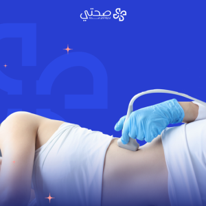
Ultrasound imaging
₺9,000What is ultrasound imaging?
Ultrasound imaging is known as a non-surgical medical procedure that uses high-frequency sound waves to create images of the internal organs of the human body, blood vessels, and tissues. The device used in imaging passes through a transducer that sends sound waves to the body. When these waves hit the tissues, they bounce back to be picked up by sensors, where they are then converted into images that the doctor can interpret.
When is ultrasound imaging performed?
- Diagnosis of heart diseases: including valve diseases, birth defects, and enlargement.
- Pregnancy assessment: to determine the age of the fetus, monitor it at different stages of growth, and assess its health.
- Vascular disease assessment: such as blood clots and hardening of the arteries.
- Abdominal disease assessment: such as the gallbladder, liver, spleen, pancreas, and kidneys.
- Tumor assessment: helps determine the size and location of the tumor and guide the needle correctly for biopsy.
- Joint assessment: to diagnose tendonitis and arthritis.
What are the benefits of ultrasound imaging?
- It does not require any surgical intervention or incisions.
- It is considered safe and does not cause any pain to the patient.
- It can be performed in a relatively short time.
- There are no risks when performed several times.
- It provides live images of organs and tissues.
What to expect when taking an ultrasound?
- Drink some water before the examination to facilitate viewing the bladder.
- Lie on the examination table.
- The doctor places a warm gel on the area to be examined.
- The doctor moves the device over the skin to take pictures.
- The doctor explains the results after the examination is complete.
-
Quick View
Ultrasound of internal parts
₺9,000What is an internal ultrasound?
An internal ultrasound is an imaging technique that uses high-frequency sound waves to create images of the body’s internal tissues and organs. It is also called “echo” or “sonar”.
What are the benefits of an internal ultrasound?
- It is an ideal option for children and pregnant women as it does not expose them to any radiation.
- It is performed without injections or punctures.
- It shows immediate results, allowing for quick medical decisions.
- It provides detailed images of internal tissues and organs.
- It can be used to evaluate many medical conditions.
When is an internal ultrasound performed?
- When there is abdominal pain: to evaluate the gallbladder, spleen, liver, pancreas and kidneys.
- When suffering from digestive system problems: to evaluate the intestines and stomach.
- When pregnant: to evaluate the growth of the fetus and monitor its health.
- When there are problems with the reproductive system: to evaluate the ovaries, uterus and prostate.
- When hips are present: to detect any tumors and determine their location and size.
- When tissue injuries are present: to evaluate tendons, muscles and ligaments.
What to expect during an internal ultrasound?
- This examination usually takes between 15-30 minutes.
- The patient is asked to undress in the examination area or wear a loose gown.
- A gel is applied to the area to be examined.
- The doctor places a probe on the skin, which sends out sound waves and receives their echo.
- The images appear on a computer screen.
- The patient may be asked to change positions during the examination in order to obtain images from different angles.
-
Quick View
X-ray imaging
₺4,500What is X-ray?
X-ray is a medical test that uses X-rays to create images of bones and other dense tissues in the body. It does not penetrate soft tissues such as organs and muscles, which is why these tissues appear dark in the images.
What are the benefits of X-ray?
- It only takes a few seconds in most cases.
- It usually does not cause any pain.
- It provides detailed and accurate images of bones.
- It can be used to examine many different parts of the body.
- It is usually cheaper than other imaging tests such as CT scans and MRIs.
When is an X-ray performed?
- When diagnosing bone fractures, even small or complex ones.
- Evaluating joint diseases, as the rays show arthritis and cartilage damage.
- Searching for and detecting infections such as bone infections.
- Diagnosing chest diseases such as lung cancer, pneumonia.
What to expect when taking an X-ray?
- Wearing a gown or removing some clothing, and it may be necessary to remove metal objects and jewelry.
- You lie on an examination table or stand in front of the imaging machine, where the technician positions your body in the correct position and sends the rays through your body. After the test, the
- patient does not experience any side effects and can return to his normal activities immediately.