
Ultrasound imaging
₺9,000
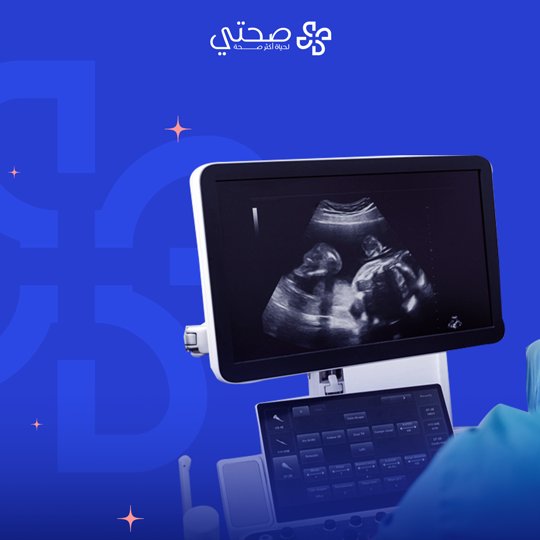
What is ultrasound imaging?
Ultrasound imaging is known as a non-surgical medical procedure that uses high-frequency sound waves to create images of the internal organs of the human body, blood vessels, and tissues. The device used in imaging passes through a transducer that sends sound waves to the body. When these waves hit the tissues, they bounce back to be picked up by sensors, where they are then converted into images that the doctor can interpret.
When is ultrasound imaging performed?
- Diagnosis of heart diseases: including valve diseases, birth defects, and enlargement.
- Pregnancy assessment: to determine the age of the fetus, monitor it at different stages of growth, and assess its health.
- Vascular disease assessment: such as blood clots and hardening of the arteries.
- Abdominal disease assessment: such as the gallbladder, liver, spleen, pancreas, and kidneys.
- Tumor assessment: helps determine the size and location of the tumor and guide the needle correctly for biopsy.
- Joint assessment: to diagnose tendonitis and arthritis.
What are the benefits of ultrasound imaging?
- It does not require any surgical intervention or incisions.
- It is considered safe and does not cause any pain to the patient.
- It can be performed in a relatively short time.
- There are no risks when performed several times.
- It provides live images of organs and tissues.
What to expect when taking an ultrasound?
- Drink some water before the examination to facilitate viewing the bladder.
- Lie on the examination table.
- The doctor places a warm gel on the area to be examined.
- The doctor moves the device over the skin to take pictures.
- The doctor explains the results after the examination is complete.
Related Products
-
Quick View
My health bouquet
₺4,550


-
Quick View
Rocketan analyzes
₺2,500