
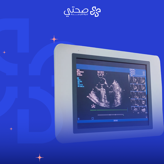
Echo imaging
₺9,000
What is an echocardiogram?
An echocardiogram is a non-invasive ultrasound test that creates moving images of the heart and its valves. These images provide accurate and valuable information about the function and structure of the heart, helping to diagnose and evaluate many heart diseases.
When is an echocardiogram performed?
- When experiencing chest pain.
- When experiencing shortness of breath.
- When experiencing fatigue.
- When experiencing leg swelling.
- When experiencing difficulty exercising.
- When experiencing a chronic cough.
- When experiencing fainting.
- When experiencing an abnormal heart sound.
What are the benefits of an echocardiogram?
- Diagnosing heart diseases: An echocardiogram detects heart diseases including: valve diseases, pericardial diseases, cardiomyopathy, congenital defects, and blood clots.
- Evaluating heart function: An echocardiogram can measure the size of the heart and reveal its pumping function, which helps to evaluate how long it is working efficiently.
- Monitoring treatment: It can be used to monitor the effectiveness of heart disease treatments, such as medications and heart valve surgery.
What to expect during an echo?
- It usually takes about 30 minutes.
- The patient is asked to lie on an examination table.
- The ultrasound technician applies gel to the patient’s chest.
- The images appear on a computer screen.
- The patient may be asked to change position or hold their breath.
Related Products


-
Quick View
Ultrasound imaging
₺9,000
